Home
Uncategories
Shoulder Joint Anatomy Diagram : Frozen Shoulder (Adhesive Capsulitis) - is it causing your Shoulder Pain? / This mobility provides the upper extremity with tremendous range of motion such as adduction, abduction, flexion, extension, internal rotation, external rotation, and 360° circumduction in the shoulder joint anatomy.
Shoulder Joint Anatomy Diagram : Frozen Shoulder (Adhesive Capsulitis) - is it causing your Shoulder Pain? / This mobility provides the upper extremity with tremendous range of motion such as adduction, abduction, flexion, extension, internal rotation, external rotation, and 360° circumduction in the shoulder joint anatomy.
Shoulder Joint Anatomy Diagram : Frozen Shoulder (Adhesive Capsulitis) - is it causing your Shoulder Pain? / This mobility provides the upper extremity with tremendous range of motion such as adduction, abduction, flexion, extension, internal rotation, external rotation, and 360° circumduction in the shoulder joint anatomy.. The shoulder joint is vulnerable to dislocations from sudden jerks of the arm, especially in children before strong muscles have developed. It stretches across the top of the shoulder from the clavicle in the front to the scapula in the back. This mobility allows you to move through a tremendous range of motion in a variety of planes. The shoulder muscles bridge the transitions from the torso. Editor · aug 6, 2017 ·.
Click now and learn everything about its anatomy and function at kenhub! The next layer is made up of the ligaments of the joint capsule. There are actually four joints within the shoulder: Dislocation of the shoulder is extremely painful and may require surgical repair or even cause permanent damage. This mobility provides the upper extremity with tremendous range of motion such as adduction, abduction, flexion, extension, internal rotation, external rotation, and 360° circumduction in the shoulder joint anatomy.
Shoulder Muscles Diagram - Muscles Of The Shoulder Joint And Girdle Human Anatomy Kenhub Youtube ... from etc.usf.edu The small size of the glenoid fossa and the relative laxity of the joint capsule renders the joint relatively unstable and prone to subluxation and. 7 draw labelled diagram showing the relations of shoulder joint. 8 name the arteries and the. Equally extensive are the muscles affecting the shoulder movement, including: The shoulder muscles bridge the transitions from the torso. The glenohumearal joint has a greater range of motion than any other joint in the body. The shoulder joint is formed where the humerus (upper arm bone) fits into the scapula (shoulder blade), like a ball and socket. Editor · aug 6, 2017 ·.
The shoulder joint comprises parts of the following bone structures:
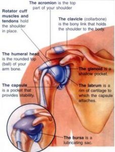
The the glenohumeral joint is what most people think of as the shoulder joint. It stretches across the top of the shoulder from the clavicle in the front to the scapula in the back. The next layer is made up of the ligaments of the joint capsule. Webmd's shoulder anatomy page provides an image of the parts of the shoulder and describes its function, shoulder problems, and more. The shoulder joint comprises parts of the following bone structures: It is the major joint connecting the upper limb to the trunk. The fixed joint capsule forms an envelope around the shoulder joint to seal it off from the surrounding tissue. The shoulder has about eight muscles that attach to the scapula, humerus, and clavicle. Normal anatomy, variants and checklist. Click here to watch an anatomy video about the shoulder joint anatomy. The shoulder joint by quan fu gan 60492 views. The small size of the glenoid fossa and the relative laxity of the joint capsule renders the joint relatively unstable and prone to subluxation and. It has the largest range of motion out of all the joints in the body and consists of three bones.
Shoulder joint of human body anatomy infographic diagram with all parts including bones ligaments muscles bursa cavity capsule cartilage membrane for medical science education and health care. Webmd's shoulder anatomy page provides an image of the parts of the shoulder and describes its function, shoulder problems, and more. 7 draw labelled diagram showing the relations of shoulder joint. The shoulder joint (glenohumeral joint) is a ball and socket joint between the scapula and the humerus. Atlas of the anatomy of the joint of the shoulder on a ct arthrogram in axial, coronal, and sagittal sections, on a 3d images and on conventional athrogram.
Shoulder Anatomy from www.fpnotebook.com The shoulder joint is vulnerable to dislocations from sudden jerks of the arm, especially in children before strong muscles have developed. The human shoulder is the most mobile joint in the body. There are actually four joints within the shoulder: The deepest layer of the shoulder includes the bones and the joints. Learn vocabulary, terms and more with flashcards, games and other study tools. The fixed joint capsule forms an envelope around the shoulder joint to seal it off from the surrounding tissue. This acts as the bony framework by which the muscles of the chest, upper back and shoulder connect the upper limb to the trunk of the body and control it's movements.the clavicle connects to the sternum via the sternoclavicular joint and to the scapula by. The shoulder has about eight muscles that attach to the scapula, humerus, and clavicle.
The glenohumearal joint has a greater range of motion than any other joint in the body.
6 describe briefly the abduction at shoulder joint. The shoulder has about eight muscles that attach to the scapula, humerus, and clavicle. The glenohumearal joint has a greater range of motion than any other joint in the body. Atlas of the anatomy of the joint of the shoulder on a ct arthrogram in axial, coronal, and sagittal sections, on a 3d images and on conventional athrogram. Learn vocabulary, terms and more with flashcards, games and other study tools. The next layer is made up of the ligaments of the joint capsule. It stretches across the top of the shoulder from the clavicle in the front to the scapula in the back. There are several types of joints including pivot, hinge, saddle and ball and socket joints. It's the major joint in the shoulder, where the rounded top, or head, of the. In this article, we shall look at the anatomy of the shoulder joint and its important clinical correlations. License image the shoulder joint ligaments shown are the acromioclavicular ligament, coracoacromial ligament, coracohumeral ligament, coracoclavicular ligament, and the articular capsule or glenohumeral. The shoulder joint is the connection between the chest and the upper extremity. Clavicle fracture with broken collarbone vector illustration.
In this article, we shall look at the anatomy of the shoulder joint and its important clinical correlations. Equally extensive are the muscles affecting the shoulder movement, including: Shoulder joint of human body anatomy infographic diagram with all parts including bones ligaments muscles bursa cavity capsule cartilage membrane for medical science education and health care. The shoulder joint by quan fu gan 60492 views. This mobility allows you to move through a tremendous range of motion in a variety of planes.
Rotator Cuff & Shoulder Pain - Advanced Sports & Family Chiropractic & Acupuncture from asfca.com The small size of the glenoid fossa and the relative laxity of the joint capsule renders the joint relatively unstable and prone to subluxation and. The head of the humerus: Three thickenings of the articular capsule over the anterior surface of the joint that plays very little role in strength and is mainly for joint stabilization. The shoulder joint is vulnerable to dislocations from sudden jerks of the arm, especially in children before strong muscles have developed. The the glenohumeral joint is what most people think of as the shoulder joint. This incongruent bony anatomy allows for the wide range of movement available at the shoulder joint but is also the reason for the lack of joint stability. The human shoulder is the most mobile joint in the body. The next layer is made up of the ligaments of the joint capsule.
The shoulder joint comprises parts of the following bone structures:
In this article, we shall look at the anatomy of the shoulder joint and its important clinical correlations. It has the largest range of motion out of all the joints in the body and consists of three bones. It is the major joint connecting the upper limb to the trunk. The shoulder joint by quan fu gan 60492 views. Shoulder joint of human body anatomy infographic diagram with all parts including bones ligaments muscles bursa cavity capsule cartilage membrane for human shoulder joint pain anatomy. Joint anatomy,how to draw elbow joint,elbow joint,shoulder joint,how to draw hinge joint,easy diagram,how to,how to draw ball and socket joint, how to draw hinge joint do like, subscribe, share and comment thanks for watching. Learn more about the shoulder joint anatomy. The shoulder joint (glenohumeral joint) is a ball and socket joint between the scapula and the humerus. The head of the humerus: The deepest layer of the shoulder includes the bones and the joints. Shoulder joint is the most mobile joint of the human body. Learn vocabulary, terms and more with flashcards, games and other study tools. Joints are the connections between bones in the human skeleton.
Shoulder joint is the most mobile joint of the human body shoulder anatomy diagram. The shoulder joint is the connection between the chest and the upper extremity.


0 Comments:
Posting Komentar